Left and right differ.
Organ layout, lung structure, and diaphragm shape are asymmetric. Asymmetry itself is not a defect.
 MOVEMENT
MWK FOUNDATIONS / ASYMMETRY & LATERALIZATION
The two sides of your body are not mirror copies. Understanding that difference is a foundation of how we observe movement at Movement with Kev.
Start with the anatomy. Use a movement model to ask better questions. Let the person’s actual movement guide the next step.
01 / THE ANATOMY
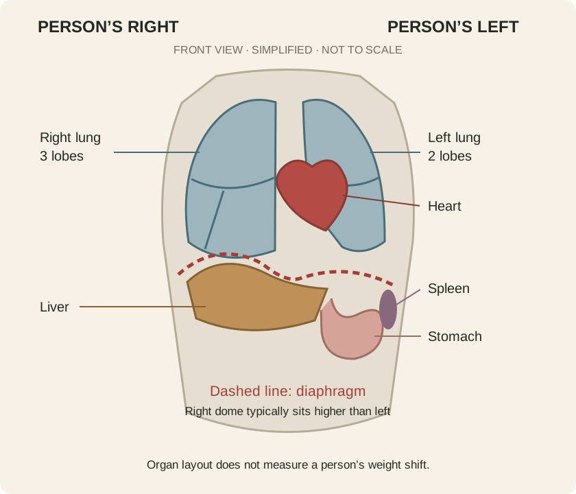
In the typical organ arrangement, most of the liver sits on the right; the stomach and spleen lie mainly on the left. The lungs have different shapes and lobe counts, with space for the heart on the left. [1, 2]
The right and left diaphragm domes also differ in position and shape. A study of 153 adults with normal pulmonary function found a higher right dome on average, alongside substantial variation between people. [3]
This diagram shows typical anatomy. Some people have reversed organ placement (situs inversus) or other arrangements. It is not an individual scan or a map of weight distribution.
02 / THE CONNECTION TO MOVEMENT
The diaphragm has respiratory and postural roles. Human experiments have measured diaphragm activity during rapid arm movement and linked coordinated diaphragm–abdominal activity to changes in intra-abdominal pressure. [4, 5]
Organ layout, lung structure, and diaphragm shape are asymmetric. Asymmetry itself is not a defect.
The diaphragm participates in breathing and postural tasks. That finding alone does not specify which leg a person loads.
We use contacts, position, pressure control, and retesting to explore movement on both sides. This is how we apply the framework.
03 / UNDERSTANDING RIGHT LATERALIZATION
In MWK’s coaching language, right lateralization describes an observed preference for organizing support toward the right. It is a working description that needs a clear task and repeatable observations.
Some movement frameworks propose that typical organ and diaphragm asymmetry contributes to a common right-sided bias. We treat that proposed connection as a model—not proof that every person shifts right because of their organs.
Organ asymmetry, handedness, and weight-bearing preference are different observations. One does not establish the others. A study of 41 healthy young adults found that weight distribution changed over time and differed between relaxed and constrained standing. [6]
Our foundation: respect natural asymmetry, assess the individual, and develop usable movement options on both sides.
We do not use organ layout alone to label a pelvis, explain pain, or choose a corrective exercise. The aim is adaptable movement—not perfectly identical sides or a permanent 50/50 stance.
04 / BRING IT INTO PRACTICE
Use the Visual Read to describe contacts and position before assigning a pattern.
Keep the setup consistent. Record what each side can do, how it feels, and where the setup first changes.
Within an appropriate practice, adjust a support, contact, or cue. The response is information—not a diagnosis or proof of an organ-based cause.
Compare comfort and available movement. Keep useful changes and reconsider the model when the observations do not fit.
EVIDENCE & FURTHER STUDY
Sources support the specific anatomy and experimental findings described above. They do not establish a universal organ-driven right-lateralized posture.
Typical organ layout and the existence of reversed arrangements.
Right–left differences in lung lobes and space occupied by the heart.
Observational imaging study; anatomy varies across individuals. It did not test a universal right weight shift.
Experimental evidence of diaphragm involvement in a postural task.
Pressure and muscle activity during experimental movement tasks.
Small healthy sample; standing strategy varied across tasks and time. Not an organ-causation study.